How TI-RADS supports thyroid ultrasound
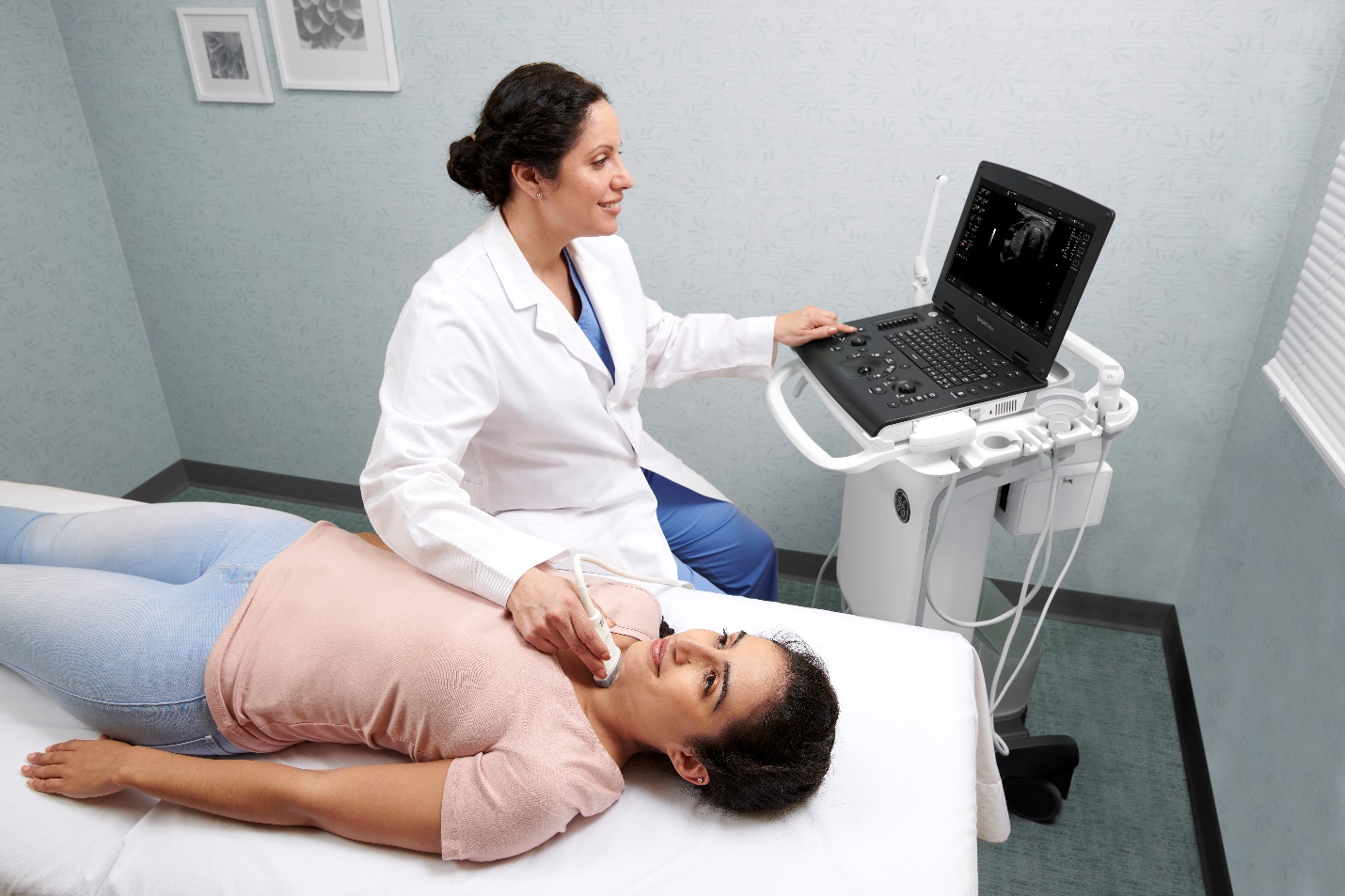
Thyroid ultrasound is an increasingly common diagnostic tool for general practitioners (GPs) to make patient care more convenient and effective.1 The ability to perform scans to identify nodules, goiters, abnormal lymph nodes, and other issues has allowed for faster and more efficient diagnosis.
One of the factors making this possible is the collective reliance on the American College of Radiology Thyroid Imaging, Reporting and Data System (ACR TI-RADS™).2
The TI-RADS system uses a standardized scoring model for reports, providing users with guidance for when to recommend ultrasound follow-up or fine needle aspiration of suspicious nodules.
What are the main benefits of TI-RADS scoring?
TI-RADS has standardized vocabulary to assist in assessing nodule malignancy risk level. It also facilitates clinical consensus and agreement among different members of patients' care teams and decreases reader variability when it comes to comparisons of scan results.3
The system has been found to be a simple and practical method of assessing thyroid nodules with high positive predictive value (PPV) and good inter-observer agreement.4 This helps ensure that all clinicians are aligned in their interpretation of results, offers better speed of care, and allows for a more proactive treatment plan.
Thyroid cancer is among the most common types in the United States and can spread rapidly,5 even while clinicians relay their difference of opinions on scan findings.
While there still may be differing opinions regarding the finality of diagnosis and a certain degree of reader variability, TI-RADS scoring significantly improves clinician agreement.6
TI-RADS scoring has also been found to help reduce unnecessary biopsies, benefiting doctors and patients alike.7 With thyroid nodules occurring in over 76% of adults and only around five percent being malignant, TI-RADS scoring, and thyroid ultrasound in general can help clinicians risk stratify so that a majority of patients with lower risk stratification are prevented from invasive, needle-focused procedures while providing maximum diagnostic accuracy and confidence.
How are TI-RADS scores calculated?
The TI-RADS scoring and risk stratification system has proven highly sensitive for detecting malignancy in thyroid nodules and is a reliable technique for the initial assessment of thyroid nodules and subsequent clinical decision-making. Clinicians can use a universal scale from 0 to 5 to detect malignancy risk, the need for fine-needle aspiration, and directives for subsequent follow-up.
- TI-RADS 1 - Benign with no need for FNA
- TI-RADS 2 - Not Suspicious (No need for FNA)
- TI-RADS 3 - Mildly Suspicious (FNA if greater than 2.5 cm and follow-up if greater than 1.5 cm)
- TI-RADS 4 - Moderately Suspicious (FNA if greater than 1.5 cm and follow-up if greater than 1 cm)
- TI-RADS 5 - Highly Suspicious (FNA if greater than 1 cm and follow-up if greater than 0.5 cm)
TI-RADS scoring relies on several distinct properties to determine malignancy risk. These include composition echogenicity (the brightness of an image caused by the reflection of sound waves), shape (taller than wide usually reflects a higher risk of malignancy), margin (extra-thyroidal extension represents the most significant likelihood of malignancy), and the presence of echogenic foci.8
How TI-RADS serves as a workflow resource for ultrasound users
TI-RADS provides a workflow resource for their reporting, diagnosis and decision-making. While thyroid ultrasound was once primarily the purview of radiologists and imaging specialists, general practitioners and internists have long been assessing nodules prior to referral.9
Now, as more and more GPs offer thyroid ultrasound in their offices,10 standardization of reporting and a user-friendly guide for interpreting scan results are essential to helping new users develop and increase confidence in their diagnosis.
This system can be especially effective in areas where specialists are in short supply or are simply unavailable. One recent study found that TI-RADS classification is an appropriate and non-invasive method for assessing thyroid nodules in routine practice and can safely reduce the number of unnecessary fine-needle aspiration in a significant proportion of benign thyroid lesions. The study findings also asserted that TI-RADS should be standardized as the screening tool in resource-limited areas.11
TI-RADS and your ultrasound system: what you need to know
As useful as TI-RADS is supporting clinicians assessment and ultrasound findings, it's important that your system has features that can quickly and accurately leverage TI-RADS reporting. The evolution of ultrasound technology has made the integration of TI-RADS reporting easier and more intuitive than ever.
Certain systems offer thyroid-specific preset packages that allow users to enter TI-RADS criteria and assessment while easily enabling labeling, measuring, and description of nodules, lymph nodes, and parathyroid. They also enable clinicians to categorize lesions found on thyroids. Users can also look at first-level diagnosis categorization to determine the need for future imaging or FNA biopsy, all through one centralized process. Comments and descriptions entered during scans are fed directly into the TI-RADS report.
Whether you're just starting to perform thyroid ultrasound in your practice or are looking for better ways to incorporate TI-RADS scoring in your scans, it's important to have the right system and processes in place so you can marry the simplicity of TI-RADS with the simplicity of your own workflow.
TI-RADS is a trademark of the American College of Radiology.
REFERENCES:
- Quianzon, C. C., & Schroeder, P. R. (2015). Initial evaluation of thyroid nodules by primary care physicians and internal medicine residents. Journal of community hospital internal medicine perspectives, 5(2), 27192. https://doi.org/10.3402/jchimp.v5.27192
- TI-RADS. (n.d.). American College of Radiology. https://www.acr.org/Clinical-Resources/Reporting-and-Data-Systems/TI-RADS
- Sahli, Z. T., Sharma, A. K., Canner, J. K., Karipineni, F., Ali, O., Kawamoto, S., Hang, J. F., Mathur, A., Ali, S. Z., Zeiger, M. A., & Sheth, S. (2019). TIRADS Interobserver Variability Among Indeterminate Thyroid Nodules: A Single-Institution Study. Journal of ultrasound in medicine: official journal of the American Institute of Ultrasound in Medicine, 38(7), 1807–1813. https://doi.org/10.1002/jum.14870
- Chandramohan, A., Khurana, A., Pushpa, B. T., Manipadam, M. T., Naik, D., Thomas, N., Abraham, D., & Paul, M. J. (2016). Is TIRADS a practical and accurate system for use in daily clinical practice? The Indian journal of radiology & imaging, 26(1), 145–152. https://doi.org/10.4103/0971-3026.178367
- American Thyroid Association. (2019, October 14). Vol 12 Issue 10 P.7-8 | American Thyroid Association. https://www.thyroid.org/patient-thyroid-information/ct-for-patients/october-2019/vol-12-issue-10-p-7-8/
- Ko, S. Y., Lee, H. S., Kim, E. K., & Kwak, J. Y. (2014). Application of the Thyroid Imaging Reporting and Data System in thyroid ultrasonography interpretation by less experienced physicians. Ultrasonography (Seoul, Korea), 33(1), 49–57. https://doi.org/10.14366/usg.13016
- Abou Shaar, B., Meteb, M., Awad El-Karim, G., & Almalki, Y. (2022). Reducing the Number of Unnecessary Thyroid Nodule Biopsies With the American College of Radiology (ACR) Thyroid Imaging Reporting and Data System (TI-RADS). Cureus, 14(3), e23118. https://doi.org/10.7759/cureus.23118
- Botz, B., & Smith, D. (2017). ACR Thyroid Imaging Reporting and Data System (ACR TI-RADS). Radiopaedia.org. https://doi.org/10.53347/rid-52374
- Anwar, K., Mohammad, A. Y., & Khan, S. (2023). The sensitivity of TIRADS scoring on ultrasonography in the management of thyroid nodules. Pakistan journal of medical sciences, 39(3), 870–874. https://doi.org/10.12669/pjms.39.3.7313
- Quianzon, C. C., & Schroeder, P. (2015). Initial evaluation of thyroid nodules by primary care physicians and internal medicine residents. Journal of Community Hospital Internal Medicine Perspectives, 5(2), 27192. https://doi.org/10.3402/jchimp.v5.27192
- Genc, A., Ryk, M., Suwała, M., Żurakowska, T., & Kosiak, W. (2016). Ultrasound imaging in the general practitioner's office - a literature review. Journal of ultrasonography, 16(64), 78–86.
- https://doi.org/10.15557/JoU.2016.0008Isse, H.M., Lukande, R., Sereke, S.G. et al. Correlation of the ultrasound thyroid imaging reporting and data system with cytology findings among patients in Uganda. Thyroid Res16, 26 (2023). https://doi.org/10.1186/s13044-023-00169-1