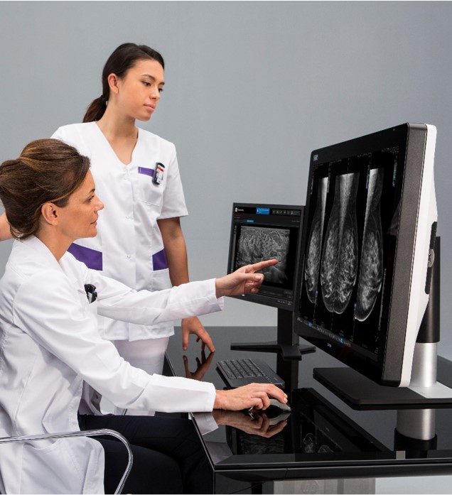
Breast cancer currently affects more than one in ten women worldwide,[1] and became the most common cancer globally as of 2021, accounting for 12 percent of all new annual cancer cases worldwide, according to the World Health Organization.[2] However, in the US, the overall death rate from breast cancer decreased by 1 percent per year from 2013 to 2018 and these decreases are thought to be the result of treatment advances such as 3D breast imaging and earlier detection through screening. Every day, advances in mammography technology such as 3D breast imaging or digital breast tomosynthesis (DBT) have a significant impact on a woman’s cancer screening experience, resulting in better image quality than traditional 2D imaging, and higher specificity and the potential for improved cancer detection. These advances have resulted in fewer false positives and the need for fewer biopsies.[3] Some 3D exams can also be acquired faster and can potentially reduce radiation exposure to patients.
The benefits of advanced imaging technology in women’s health not only support the patient experience but also contribute to an optimized workflow for the clinicians and technologists performing the exams. Industry leaders in women’s health imaging, such as GE Healthcare, are also improving other facets of the imaging workflow to not only help support a positive patient experience, but also create a more efficient experience for the mammography technologist, physicist, and other radiology staff.
Upgrading efficiency in the quality control process
Optimized clinical workflows in mammography operations are necessary to support the large patient volumes of annual screening mammogram visits. Over 42 million screening mammograms are performed each year in the US and UK, but despite that number, many women are not getting their screenings.[4] As hospitals and women’s health centers continue efforts to improve participation in annual screening programs, imaging manufacturers such as GE Healthcare are working to incorporate intelligent efficiencies to 3D breast imaging and biopsy workflows to support potentially higher volumes of screening mammograms and follow-up care.
As a result, routine performance testing of mammography systems is necessary to assess clinical performance indicators and ensure the production of high-quality images, with the least possible amount of radiation exposure that will allow the radiologist to accurately detect breast cancer or other breast pathologies such as fibrosis and simple cysts, intraductal papillomas or duct ectasia, among others. These manual quality control checks can be time consuming for mammography technologists and can take time from patient exams.
Automating the quality control (QC) protocols, which subsequently requires much less manual effort by the mammography technologist, and faster completion of the process at each necessary interval, is critical for mammography systems like the Senographe Pristina™. This single automated process can help save time for the mammography technologists, especially when there are several systems that require performance testing.
“Using the new automated QC process, I’ve been able to run through the protocols in about half the time as I would need before."
--Meggan Bush, Radiology Technologist at Lake Medical Imaging
According to Meggan Bush, Radiology Technologist at Lake Medical Imaging in The Villages, Florida, the time savings she has experienced using Pristina’s automated QC process has been significant. “I’ve been doing the quality control here for about nine years,” she explained. “Using the new automated QC process, I’ve been able to run through the protocols in about half the time as I would need before. There’s no running back and forth to reset the machine and run another set of images. And it’s reassuring to know that as an automated process, we’re ensuring that our systems are producing the same quality imaging for every patient.”
At Lake Medical Imaging, the technologists are performing about 350 mammograms per week. Another facility, Hopital Pierre-le Gardeur in Terrebonne, Quebec, Canada, has also installed the Pristina quality control upgrade. Staff spend about fifteen minutes per system to complete QC protocols on their three different mammography systems, showing a significant savings. Hopital Pierre-le Gardeur does not have one dedicated QC technologist but requires all mammography technologists to perform QC protocols.
“The feedback we’ve received over the last six months,” explained Marie-Claude L’Esperance, MD, Radiologist at Hopital Pierre-le Gardeur, “has been really positive. The improvements remove all the manual work and adjusting that has been necessary to perform our QC protocols. It’s a lot faster.”
The upgraded quality control feature automates the daily and monthly protocols, simplifying the process, which can be especially important for new or less experienced staff, according to the Quebec team.
“Our technologists now have more certainty completing the QC protocols. We’re finding there’s less need for repeating tests."
--Denise Robichaud, Radiology Technologist at Hopital Pierre-le Gardeur
The team’s increased confidence also flows into patient care, L’Esperance noted. She explained that the mammography technologists are more in control and feel more comfortable and confident. And now that they have less to do, they can spend more time with the patient.
Optimizing the biopsy workflow with sample imaging
At the Quebec facility, the biopsy workflow used to be a cumbersome process for both the patient as well as the staff. Using a legacy system, the Quebec team performed biopsies as patients lay prone on the exam table, which limited patient comfort, extended the procedure time, and sometimes caused patients additional anxiety, according to the clinical team at the facility.
Additionally, the team explained, no screening mammograms could be scheduled in the room adjacent to the biopsy room during the procedure. Once a tissue sample was taken, the specimen would be imaged on the mammography system in the adjacent room.
When the Quebec facility updated their women’s health imaging facilities with 3D mammography, it also invested in two of GE Healthcare’s Pristina Serena™ biopsy systems, which has significantly optimized both the clinical workflow and scheduling.
“In terms of workflow,” L’Esperance said, “we are now able to image the tissue specimen right there, without having to block an additional room to image the biopsy samples. It was that final piece of the puzzle for us. We have truly elevated our patient care. We have invested in 3D mammography for the benefit of our patients for cancer detection, and now biopsies can be performed while the patients are sitting up. The procedure is faster and it’s a lot less traumatizing for the patients who used to have to lay on their stomachs for an hour. They are much more comfortable now, and our radiologists love the image quality they’re getting from all of the investments we’ve made in our breast care facility.”
Advances in women’s health imaging technology, from imaging acquisition to workflow, are helping to improve earlier detection of breast cancer in annual mammography screening, and also improve the imaging and care experience, for both patients and staff.
To learn more about the efficiency suite offerings for quality check as well as sample imaging, please visit GE Healthcare’s Senographe Pristina and Pristina Serena.
The results achieved by these facilities may not be applicable to all institutions and individual results may vary. This is provided for informational purposes only and its content does not constitute a representation or guarantee from GE Healthcare.
Not all products or features are available in all geographies. Check with your local GE Healthcare representative for availability in your country.
1 Yedjou CG, Sims JN, Miele L, et al. Health and Racial Disparity in Breast Cancer. Adv Exp Med Biol. 2019;1152:31-49. doi:10.1007/978-3-030-20301-6_3. https://www.ncbi.nlm.nih.gov/pmc/articles/PMC6941147/
[2] https://www.breastcancer.org/symptoms/understand_bc/statistics


