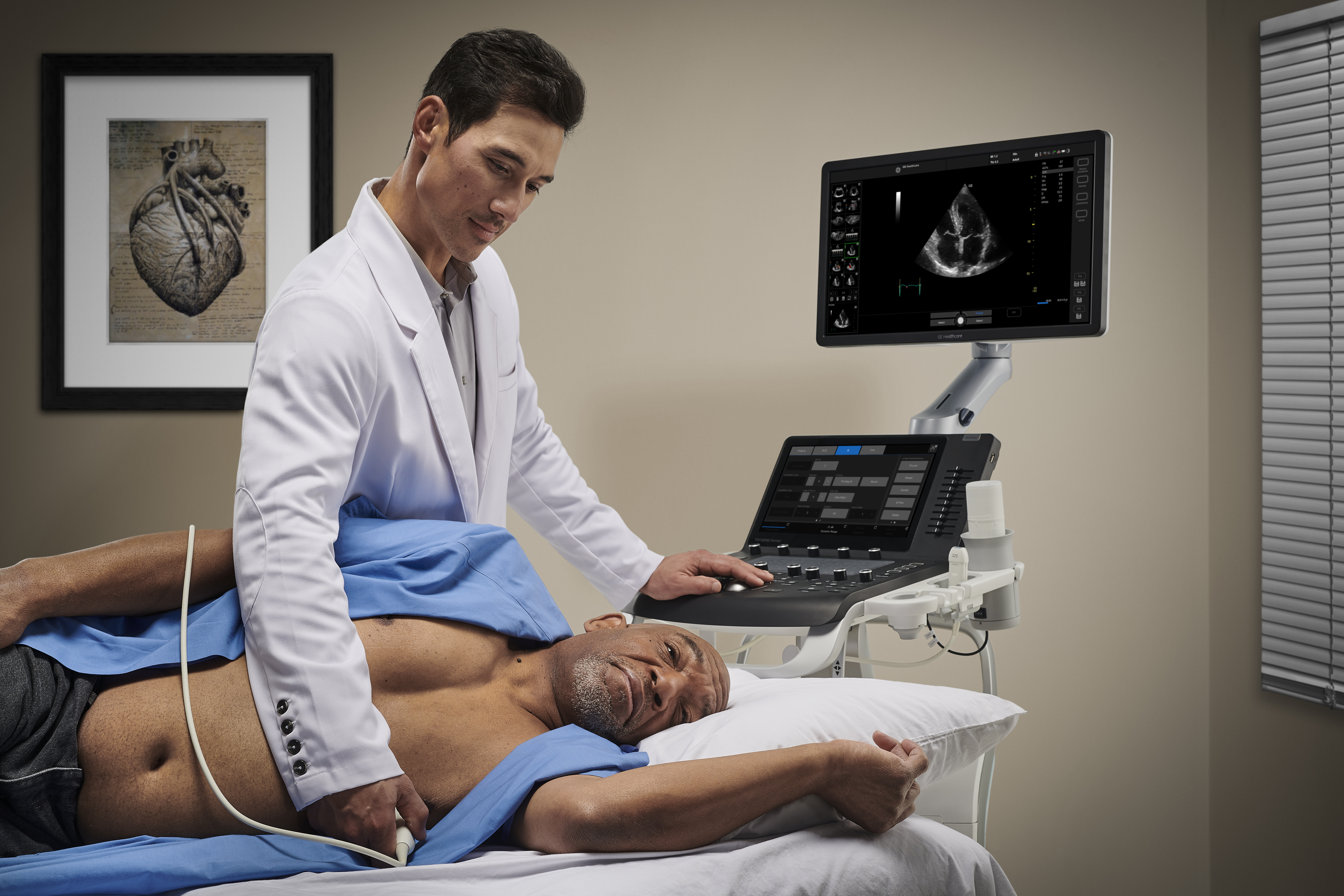
Cardiac care is among the many areas of specialization being brought under general practitioners' (GPs') services through the use of cardiovascular ultrasound. For years, integrating ultrasound systems into these environments has helped expedite cardiac diagnoses, alter the trajectory of care, and improve treatment outcomes for patients experiencing multiple types of heart issues.1
Cardiac ultrasound in primary care has helped improve the health and quality of life of at-risk people of all age groups.2 Cardiovascular ultrasound has often been a catalyst for midstream change-of-treatment plans based on scan findings, allowing physicians to guide patients toward the best possible course of action.
In addition to optimizing care for their patients, the integration of cardiac ultrasound in their offices has allowed general practitioners and other clinicians in the primary care landscape to grow their practices, better centralize treatment and diagnostics under one roof, and create new avenues for revenue.
In the past, physicians were often concerned with steep learning curves, financial issues, and other perceived barriers. Today's cardiac ultrasound systems offer unprecedented ease of use, affordability, and image quality to help your practice quickly integrate them into your scope of services. Let's take a look at how primary care physicians can put ultrasound on their menus.
Managing heart disease and cardiac health in primary care
No matter what type of heart issue a patient may be experiencing, whether congenital, age-related, or lifestyle-related, their primary care physician is often the first line of defense in the management of their diagnosis. This management is complex, with most interactions actually occurring in primary care offices.3 Despite the crucial role that GPs play in management, diagnosis, and ongoing monitoring, these linchpins of treatment plans are usually ceded to specialists because of the specialized nature of the ultrasound process.4
Despite the proven benefits and the ease of use with which ultrasound can now be adapted, only a fraction of GPs have started offering any type of ultrasound in their practices. That number shrinks even more within the specialized realm of cardiac care.5
In the meantime, these doctors can't get a real-time look at critical information revealed during a cardiovascular ultrasound, such as wall thickness, valvular health, ejection fraction, and others.
At a time when heart failure mortality rates have increased consistently over the past decade,6 and progression can occur as soon as weeks from diagnosis, GPs have the power to potentially help identify conditions much earlier and help their patients increase quantity and quality of life. Imagine being able to provide time-sensitive and potentially lifesaving insights to your patients without having to wait for referral feedback .
Early and often: Ultrasound's lifetime role in detecting and managing cardiac conditions
When we examine the full spectrum of people who experience cardiac health issues, it's clear that almost all subsets of patients benefit from having ultrasound in their doctors' offices.
Before birth
Let's start with the beginning of life—when a fetus is still in the womb—where ultrasound is one of the primary diagnostic mechanisms used to detect congenital heart disease (CHD).7
GPs are taking on more and more fundamental ultrasound functions,8 such as assessing weight, heart rate, gestational age-related size disparities, polyhydramnios, oligohydramnios, cervical insufficiency, gastroschisis, placenta previa, limb-size anomalies, and others.
They can also confidently add simple cardiac views to their scope of ultrasound services to further ensure fetal health. Easy-to-learn views, such as four-chamber, outflow-tract, and great-vessel views, will increase the detection of major fetal cardiac malformations.
Having these diagnoses early is essential for successful treatment. After the child is born, and even after they receive surgery to repair the defect, ultrasound is often used to monitor heart health and activity.
Later in life
The value and benefits of primary care cardiac ultrasound persist, and, in fact, are magnified as patients get older. The risk for heart disease is highest among men older than 45 and women who have been through menopause.9 Primary care ultrasound can be used to identify and monitor many of the factors that can contribute to heart disease and failure, including left ventricular hypertrophy (LVH) and valve insufficiency.
Leveraging ultrasound in LVH and other cardiac diagnoses
LVH can be a primary indicator of heart disease. It's often associated with hypertension, obesity, advanced age, and other risk factors. The reality of LVH, however, is that patients often don't show symptoms until LVH manifests in a more serious heart condition.
Ultrasound at the point of care has long been used to detect the presence of cardiac conditions and help patients take preventative measures against different types of heart disease.10 General practitioners can now leverage this diagnostic resource to help their patients avoid the escalation of adverse cardiac health outcomes.
Primary care ultrasound can also be effective in the detection of different types of valve insufficiency and/or aortic stenosis, which are common valve issues requiring surgery but are often delayed in diagnosis.11 Ultrasound has been proven to help clinicians fine-tune their treatment approach, at times even compelling them to switch course completely based on scan results.12 When it comes to time-sensitive and extremely urgent cardiac conditions, this can be a game-changing resource in care.
Integrating cardiac ultrasound into your practice
While there have been multiple barriers to the successful integration of cardiac ultrasound in primary care, many of them are becoming increasingly navigable, including the learning curve and economic costs. In the United States, heart disease is most common in rural areas, but many primary care practices lack the resources to invest in a system. The same can be said globally for economically disadvantaged where heart disease is high, and healthcare budgets are low.13
The good news is current ultrasound systems have made it easier than ever for GPs to effectively and affordably leverage ultrasound to manage their heart disease, no matter where they are in their journey. Multiple exam protocols, tools, and applications can help you diagnose a wide spectrum of cardiovascular conditions accurately and efficiently.
Features like automated image data optimization and spectral data analysis enable clearer-than-ever scans, while intuitive measuring tools allow for quick size-referencing of heart anatomy.
At the same time, customizable scan protocols and presets facilitate ease and speed of scanning. Follow-up tools allow users to accurately perform consecutive scans and compare a previous ultrasound exam during live scanning of anomalies for accurate progression diagnosis.
To help GPs navigate inevitable learning curves, the best systems have onboarding training tools for easy system setup, operation, cleaning, and maintenance. They also offer robust training and education from vendor personnel so you and your staff can maximize operational proficiency.
Additionally, more affordable price tags and flexible financing agreements allow primary care physicians to make a smaller initial investment in ultrasound so they can quickly start reaping its growth benefits and performing scans with the highest level of confidence. Longer battery life, next-level maneuverability, and faster boot-up times have allowed ultrasound systems to thrive in all primary care environments.
Whether you're caring for patients at the beginning or toward the end of life, or simply want to offer more comprehensive services for early detection of cardiac issues, current advancements are making it simpler. More intuitive tech, lower initial costs, and comprehensive training and support have made it considerably easier to put cardiac ultrasound at the heart of your practice.
Learn more about how primary care ultrasound systems can enhance cardiac care for general practitioners.
REFERENCES:
-
Yates J, Royse CF, Royse C, et al. Focused cardiac ultrasound is feasible in the general practice setting and alters diagnosis and management of cardiac disease. Echo Research & Practice. 2016;3(3):63–69. https://doi.org/10.1530/ERP-16-0026.
-
Leviter J, Chen L, O'Marr J, et al. The feasibility of using point-of-care ultrasound during cardiac arrest in children. Pediatric Emergency Care. 2022;39(5):347–350. https://doi.org/10.1097/pec.0000000000002741.
-
Hussey AJ, McKelvie RS, Ferrone M, et al. Primary care-based integrated disease management for heart failure: a study protocol for a cluster randomized controlled trial. BMJ Open, 2022;12(5):e058608. https://doi.org/10.1136/bmjopen-2021-058608.
-
Echocardiogram (echo). American Heart Association. May 3, 2023. https://www.heart.org/en/health-topics/heart-attack/diagnosing-a-heart-attack/echocardiogram-echo.
-
Niblock F, Byun H, Jabbarpour Y. Point-of-care ultrasound use by primary care physicians. Journal of the American Board of Family Medicine. 2021;34(4):859–860. https://doi.org/10.3122/jabfm.2021.04.200619.
-
Cardiovascular deaths saw steep rise in U.S. during first year of the COVID-19 pandemic. (2023, May 23). www.heart.org. https://www.heart.org/en/news/2023/01/25/cardiovascular-deaths-saw-steep-rise-in-us-during-first-year-of-the-covid-19-pandemic#:~:text=Cardiovascular%20disease%2Drelated%20deaths%20jumped,high%20of%20910%2C000%20in%202003
-
Sun HY. Prenatal diagnosis of congenital heart defects: echocardiography. Translational Pediatrics. 2021;10(8):2210–2224. https://doi.org/10.21037/tp-20-164.
-
Perinatal ultrasound examination. (n.d.). AAFP. https://www.aafp.org/about/policies/all/obstetric-ultrasound.html#:~:text=Pregnancy%20and%20perinatal%20care%20are,therapeutic%20capabilities%20of%20family%20physicians.
-
ABCs of Knowing your heart Risk. (2022, December 20). Johns Hopkins Medicine. https://www.hopkinsmedicine.org/health/wellness-and-prevention/abcs-of-knowing-your-heart-risk#:~:text=Men%20older%20than%20age%2045,you%20should%20be%20aware%20of.
-
Bornstein AB, Rao SS, Marwaha, K. Left ventricular hypertrophy. StatPearls. August 8, 2023. https://www.ncbi.nlm.nih.gov/books/NBK557534/.
-
Potter A, Pearce K, Hilmy N. The benefits of echocardiography in primary care. British Journal of General Practice. 2019;69(684):358–359. https://doi.org/10.3399/bjgp19x704513.
-
Weile J, Frederiksen CA, Laursen CB, et al. Point-of-care ultrasound induced changes in management of unselected patients in the emergency department - a prospective single-blinded observational trial. Scandinavian Journal of Trauma, Resuscitation and Emergency Medicine. 2020;28(1): 47. https://doi.org/10.1186/s13049-020-00740-x.
-
Risk of developing heart failure much higher in rural areas vs. urban. National Institutes of Health (NIH). January 25, 2023. https://www.nih.gov/news-events/news-releases/risk-developing-heart-failure-much-higher-rural-areas-vs-urban#:~:text=Adults%20living%20in%20rural%20areas,the%20National%20Institutes%20of%20Health.
JB30840XX

